Anatomy can be a doosey for everyone, so don't let it get you down if you're having trouble. A few mnemonics and acronyms can be your savior if you are really struggling. Just make sure you have them straight and practice saying them out loud. Remember that sometimes, the most silly or inappropriate mnemonics or acronyms can help you recall the toughest things! Be creative and have fun with anatomy, it will help it stick. Some of my acronyms and mnemonics may not be helpful or useful to you if you don't understand them, so try making your own. When in doubt, draw it out! Drawing vasculature or nerves from beginning to end can take some time, but can be worth it in the long run. Typically, I would draw out sections of vasculature or nerves and then go to the lab to identify them. It really helped me solidify relationships.
When I was in anatomy, I would read the lab manual the night before, taking notes on things I thought were important for the lecture test. To my surprise, much of what I originally thought were minor details, were actually very helpful in locating structures for the lab exam. Knowing relationships between structures can aid your learning in anatomy, because if you can find landmarks like nerves, vessels, or bony structures, you can often figure out what something is.
For your practical, if your school offers any old exams, you might make a list of all of the terms that have been tested on in the past and their occurrence. You and your classmates might also join Quizlet and start your own flashcard deck. Our class created decks for each exam that helped to quiz when the lab was closed. Creating tables with muscle origins, insertions, and actions will also help reinforce your learning. Remember, teamwork makes the dream work!
If you have anything to add to this list, please leave it in the comments section below. Thank you all for reading! Much thanks to the UT Southwestern PA Class of 2015 for their help.
Helpful Links
- The Skeleton Dance - for help with bony landmarks
Upper Body
- Suprascapular nerve/artery with the superior transverse scapular ligament (bridge)
- Army goes over the bridge, Navy goes under it
- SITS: Supraspinatus, Infraspinatus, Teres MINOR, Subscapularis
- SIT attaches to greater tubercle
- Subscapularis attaches to lesser tubercle
- First S = abductor
- IT = lateral rotators
- Last S = medial rotator
- Brachial Plexus
- Real Texans Drink Cold Beer => Roots, Trunks, Divisions, Cords, Branches
- Branches of Posterior Cord: ULTRA
- Upper subscapular
- Lower subscapular
- Thoracodosral
- Radial
- Axillary
- Axillary Artery Branches
- Screw the Lawyer, Save A Patient
- Superior thoracic artery
- Thoraco-acromial artery
- Lateral thoracic artery
- Subscapular artery
- Anterior circumflex humeral artery
- Posterior circumflex humeral artery
- C5,6,7 Raise your wings to heaven--> C5, C6, C7 contributions form the long thoracic nerve, which innervates the serratus anterior. One of the actions of the serratus anterior is upward rotation of the scapula as you lift your arms at the glenohumeral joint. You can also say C5,6,7 keep your wings from heaven, as an injury to the long thoracic nerve will cause winging of the scapula
- Movements of the Pectoral Girdle (Scapula)
- Depression - "People Like Sex After Tacos"
- Pectoralis major & minor
- Latissimus dorsi
- Serratus Anterior
- Trapezius
- Protraction (PEC-S)
- Pectoralis major
- Pectoralis minor
- Serratus anterior
- Retraction - TRL (like the old MTV show)
- Trapezius (middle)
- Rhomboids (major/minor)
- Latissimus dorsi
- Superior Rotation (TrapS)
- Trapezius (superior, inferior)
- Serratus anterior (anterior, inferior)
- Inferior Rotation (PiLLaR)
- Pectoralis major (inferior, sternocostal) and minor
- Latissimus dorsi
- Levator scapulae
- Rhomboids
- Movements of the Shoulder (Glenohumeral Joint)
- Adduction - PTL (Praise The Lord)
- Pectoralis major
- Teres major
- Latissimus dorsi
- Lateral rotation - TIP
- Teres minor
- Infraspinatus
- Posterior deltoid
- Medial Rotation - SPLAT
- Subscapularis
- Pectoralis major
- Latissimus dorsi
- Anterior deltoid
- Teres major
- Contents of the Cubital Fossa (medial to lateral)
- N - median Nerve
- A - brachial Artery
- T - Tendon of the biceps brachii
- Bones of the Hand
- Looking at the right palm of hand (lateral to medial)
- Proximal row: Some Lovers Try Positions (Scaphoid, Lunate, Triquetrum, Pisiform)
- Distal row: That They Can't Handle (Trapezium, Trapezoid, Capitate, Hamate)
- LOAF Muscles of the Hand - innervated by Median Nerve
- Lateral 2 lumbricals
- Opponens pollicis
- Abductor pollicis brevis
- Flexor pollicis brevis
- Intrinsic Muscles of the Hand from Lateral to Medial
- All For One And One For All
- Abductor Pollicis Brevis
- Flexor Pollicis Brevis
- Opponens Pollicis
- Adductor Pollicis
- Opponens Digiti Minimi
- Flexor Digiti Minimi
- Abductor Digiti MInimi
- Interossei Muscles
- PAD and DAB
- Palmar interossei ADduct
- Dorsal interossei ABduct
- Contents of the Deltopectoral Triangle - CAP
- Clavicle
- Anterior deltoid
- Pectoralis major (clavicular head)
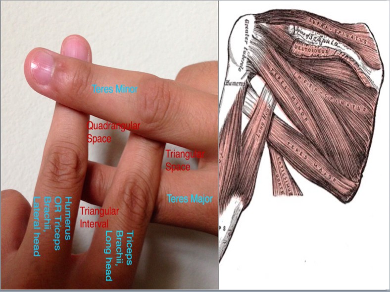
- Triangular Interval, Space, and Quadrangular Space - Finger Trick!
- Triangular Interval Boundaries - contains radial nerve and profunda brachii artery
- Long head
- Lateral head
- Teres major
- Triangular Space Boundaries - contains circumflex scapular artery
- Teres minor
- Teres major
- Long head
- Quadrangular Space Boundaries - contains axillary nerve and posterior humeral circumflex artery
- Teres minor
- Teres major
- Humerus/lateral head
- Triceps brachii (long head)
Head and Neck
- SCALP- layers of the scalp are
- Skin
- Connective tissue
- Aponeurosis
- Loose areolar tissue
- Pericranium
- Standing Room Only- The branches of CN V (in order of V1 to V3) go through the
- Superior orbital fissure
- foramen Rotundum
- foramen Ovale
- Teeth: 2123 → 2 incisors, 1 canine, 2 premolars, 3 molars (sometimes there’s only 2 molars b/c the person had no wisdom teeth)
- Cranial nerves: Oh, Oh, Oh, To Touch And Feel a Virgin Girl's Vagina And Hymen
- V3 Innervates:
- 2 pterygoids (medial and lateral)
- 2 mouth CLOSERS (temporalis and masseter)
- 2 mouth OPENERS (anterior belly of digastric, mylohyoid)
- 2 tensors (tensor veli palatini, tensor tympani)
- The branches of V1 in the superior view of the orbit from medial to lateral are NFL-- Nasocilliary, Frontal, and Lacrimal
- SO4 LR6 → Superior oblique is innervated by CN 4, Lateral Rectus is innervated by CN 6
- Actions of the Superior Oblique = 'SOLID'
- Superior Oblique Lateral rotation (Abduction) Intorsion Depression
- All of the "Glossus" muscles (genioglossus, hyoglossus, and syloglossus) are innervated by CN 12 (hypoglossal) except for the palatoglossal muscle (innervated by pharyngeal plexus)
- All of the pharyngeal muscles are innervated by the pharyngeal plexus, except for the stylopharyngeus muscle, innervated by CN 9 (glossopharyngeal)
- If the eye in question is moving toward the midline and is centered, medial rectus is working. If it moves laterally and is centered, it is lateral rectus. When medial and moving down, the superior oblique is in control. When medial and moving up, the inferior oblique is in control. Just remember that when medial, OBLIQUES are in control and they have opposite functions of their names (i.e. superior moves the eye down), whereas RECTUS (superior/inferior) muscles are in control when the eye is lateral.
- Sinuses draining into the middle meatus: AMFM → Anterior and Middle ethmoidal sinuses, Frontal sinus, and Maxillary sinus
- VAN - the order of vasculature/nerves in the intercostal space, inferior to the rib
- Vein
- Artery
- Nerve
- RALS - pulmonary artery location with respect to pulmonary bronchi in the heart
- Right - Anterior
- Left - Superior
- C3, 4, and 5 keep the diaphragm alive (phrenic nerve)
- I ate 10 Eggs At noon (foramen/hiatus locations)
- IVC - T8
- Esophageal - T10
- Aortic - T12
- The “Birds of the Thorax” or a “Duck Between 2 Gooses” in the Posterior Mediastinum
- EsophaGOOSE (Esophagus)
- Thoracic DUCK (throacic duct)
- AzyGOOSE (Azygous)
Lower Body
- External oblique = "hands in pocket" orientation
- Above the umbilicus, the artery under the rectus abdominis is the superior epigastric. Below the umbilicus, it is the inferior epigastric artery
- Abdominal retroperitoneal structures:
- SAD PUCKER
- Suprarenal glands
- Aorta/IVC
- Duodenum (2nd and 3rd part)
- Pancreas
- Ureters
- Colon (ascending/descending)
- Kidneys
- Esophagus
- Rectum
- The vas deferens passes OVER the ureters. Likewise, the uterine artery passes superior to the ureter and the vaginal artery passes inferior to the ureter (from top to bottom = UV). No inferior vesicle artery in females.
- The Pes Anserinus is made of 3 muscles and a mnemonic to remember them is "Say Grace at Tea". They are innervated by 3 different nerves and come from 3 different compartments.
- Sartorius
- Gracilis
- semiTendinosus
- The ischiocondylar (hamstring) portion of adductor magnus is more medial. Pubofemoral portion is more lateral. Together, they create the adductor hiatus.
- Gamellus muscles are innervated by the nerves innervating the muscles below them:
- Superior gamellus - nerve to obturator internus
- Obturator internus - nerve to obturator internus
- Inferior gamellus - nerve to quadratus femoris
- Quadratus femoris - nerve to quadratus femoris
- For the ligaments in the knee joint, take your middle finger on your right hand and cross it over your index finger. This represents the ligaments in your right knee, with ACL being on top, going from a posterolateral to an anteromedial position. The PCL is on bottom, going from a posteromedial to an anterolateral position. Do it with your left hand to mimic the ligaments of the left knee. Ask someone to show you this if it’s confusing.
- Movements at the Hip
- Flexors: STRIP
- Sartorius
- Tensor fascia latae
- Rectus femoris
- Iliopsoas (iliacus and psoas major)
- Pectineus
- Extensors: BASiS (I is for inferior gluteal nerve that innervates gluteus maximus, the only muscle it innervates)
- Biceps femoris (long head)
- Adductor magnus (ischiocondylar/hamstring portion)
- Semimembranosus
- Inferior gluteal nerve (gluteus maximus)
- Semitendinosus
- Lateral Rotators: GOSSIP-Q (O is for both obturators)
- Gluteus maximus
- Obturator externus/internus
- Sartorius
- Superior gamellus
- Inferior gamellus
- Piriformis
- Quadratus femoris
- Adductors: GAAAP
- Gracilis
- Adductor brevis
- Adductor longus
- Adductor magnus
- Pectineus
- Contents of the Femoral Triangle (lateral to medial) - NAVL
- femoral Nerve
- femoral Artery
- femoral Vein
- inguinal Lymphatics
- 10 named branches of internal iliac artery in the male, 11 in female
- 7 leave the pelvis
- 2 leave pelvis by crossing the pelvic brim
- Posteriorly - iliolumbar artery
- Anteriorly - umbilical artery
- Obturator canal - obturator artery
- Ventral sacral foramina - lateral sacral artery
- Greater sciatic foramen to gluteal region
- 2 pass between roots of sacral plexus
- Superior to piriformis - superior gluteal artery
- Inferior to piriformis - inferior gluteal artery
- Not passing between roots of sacral plexus
- Under coccygeus muscle, inferior to piriformis - internal pudendal artery
- 3 remaining - supply structures INSIDE the pelvis
- Superior bladder - superior vesicle artery
- Posterior bladder and prostate (male only) - inferior vesicle artery
- Wall of rectum - middle rectal artery
- branches from internal pudendal artery
- Uterus (female only) - uterine artery
- Base of bladder (female only) - vaginal artery
- LAFF muscles - innervated by the medial plantar nerve
- first Lumbrical
- Abductor hallucis
- Flexor hallucis brevis
- Flexor digitorum brevis
- Posterior Compartment of Leg
- Above Medial Malleolus
- Dick, Tom, And Very Nervous Harry (flexor Digitorum longus, Tibialis posterior, posterior tibial Artery, posterior tibial Vein, tibial Nerve, flexor Hallucis longus)
- Below Medial Malleolus
- Tom, Dick, and Very Nervous Harry (Tibialis posterior, flexor Digitorum longus, posterior tibial Artery, posterior tibial Vein, tibial Nerve, flexor Hallucis longus)
- Actions of the (Right) Leg

Credits:
- Andersenpa of physicianassistantforum.com
- UTSW Class of 2015
So just a friendly fyi, most of this material was actually compiled/created by medical students from the Class of 2015 while they were TAing the PA and PT students. Many of these mnemonics and tips were handed down to them from prior years as well. I'm glad you found them helpful!
ReplyDeleteWhoops! I was given most of these from a group of upper class PA students and I just assumed the document was created by them. Bad assumption! Thanks for clarifying that.
DeleteI went to school in Michigan and we have these same things too so pretty funny this person thinks that their class is the one who created all of these. This stuff is everywhere but having it compiled and posted in such a neat and organized way is very helpful. Thank you!
DeleteActually, pretty much all of this material has been around for decades. To suggest they were uniquely created or compiled by a class of 2015 at a medical school ascends new heights in the pursuit of hubris.
ReplyDeleteI feel like there is a lot of hatred about where the actual contents came from. I completely see your point - they were probably created well before - but why do you feel the need to argue their point or origin so much? Clearly it bothers you, but you are posting yourself as anonymous, so I feel no need to change anything because this is a public blog and I am allowed to post whatever I like because I own it.
DeleteYes this stuff has been around for ages but thank you for putting it all together so neatly. Its always easy to criticise. There are few 'doers' in this world so hats off to you. Nice to see someone passionate about what they are learning and what has made the journey easier. All the best in your studies.
ReplyDeleteThanks anonymous! I appreciate the feedback!
DeleteThank you so much for this! Extremely helpful while studying!
ReplyDeleteThank you for your positive feedback!
DeleteI personally think it is very kind of you Paul to post it for all to benefit from. Thank you
ReplyDeleteThanks for reading Eden!
DeleteThis is a great resource, thank you! I'm so glad I found your blog.
ReplyDeleteI cannot believe I found this! You are the man Paul! Lots of love from Pakistan!
ReplyDeletethanks you dear.. its really helpful. god bless you .. and lots of love for sharing this..:)
ReplyDeleteThanks for reading and for your support!
DeleteThank you for sharing this
ReplyDeleteI'm a medical student from thailand
really thanks : )
Thanks for reading and for your support!
Deletei found this helpful.thanks...medical student from Zambia
ReplyDeleteHi Paul,
ReplyDeleteGreat list of anatomy resources! I am wondering if you might be interested in adding Lecturio as well?
We provide a free medical library with lots of anatomy topic reviews here: https://www.lecturio.com/magazine/preclinical/anatomy/
We would love to hear what you think of it! :)
Paul, seriously you rock! This site has come in handy so many times and is so helpful for rotations! I have shared it with nearly all my classmates. You are the kind of PA I want to be someday. :) Thank you.
ReplyDeleteThanks so much for reading and your kind words!! I appreciate you sharing with your class. Best of luck in PA school!
DeleteEven as a paramedic finishing his last year of my B.S. this helped a lot. Thank you sir.
ReplyDeleteI am good resume writer. I am professional in resume writing. I have written various types of administrative assistant resume. In that I focus on almost all fields such as accounting, arts, clerks, and many more. medical resume template
ReplyDeleteAnother great one I have learnt is for remembering the hand bones:
ReplyDeleteA TRAPEZOID was on the TRAPEZIUM and broke his SCAPHOID and was deCAPITATEd at night (LUNATE), his mate (HAMATE) was pissing (PISIFORM) himself 3-4 times (TRIQUETRAL).